Schedule a preventative skin cancer screening and treatment at DermaBlue at our Asheville or Hendesonville locations.
Skin cancer is highly treatable when detected early. It is important to regularly examine your skin to see if any spots or moles on the skin are changing, itching, or bleeding. If you have an increased risk or family history of melanoma or skin cancer, you should be sure to have yearly skin checks by a dermatology professional.
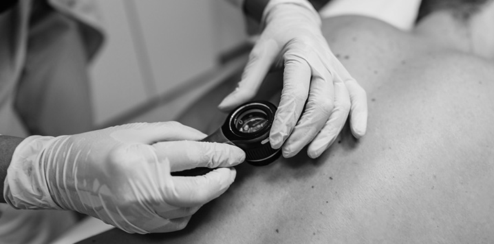
The screening with take around 10-15 minutes or longer, depending on whether or not the provider sees any unusual lesions. You will wear a medical gown and the provider will then inspect the skin including the scalp, hands, and feet. The provider will be looking for moles that show signs of cancer such as:
Asymmetry:
Moles or spots are not the same shape on both sides.
Border Irregularity:
Moles and spots have ragged, burred edges.
Color:
Moles contain different shades of tan, brown, or black.
Diameter — moles that grow larger than ¼ inch.
Evolving:
Any spot that changes over time could be a sign of skin cancer
Once the skin has been checked, the doctor will review any findings and let you know if any additional follow up is needed. Any concerning lesions will be biopsied and we will call you in a few days to review the findings. If there are additional treatments you will be scheduled to come into the office.
If we do discover skin cancer early enough, there are some in-office treatments we can perform. Additional treatment for skin cancer can include:
After a local anesthetic is injected, a surgical knife (scalpel) is used to shave off the growth stitches are not needed. Any bleeding can usually be controlled with a chemical that stops bleeding and by applying pressure. The biopsy site is then covered with a bandage or sterile dressing.
After a local anesthetic is injected, a small, sharp tool that looks like a cookie cutter called a “punch” is placed over the lesion, pushed down, and slowly rotated to remove a circular piece of skin. The skin sample is lifted up with a tool called a “forceps” or a needle and is cut from the tissue below. Stitches may not be needed for a small skin sample. If a large skin sample is taken, one or two stitches may be needed. Pressure is applied to the site until the bleeding stops. The wound is then covered with a bandage or sterile dressing.
After a local anesthetic is injected, the entire lesion is removed with a scalpel. Stitches are used to close the wound. Pressure is applied to the site until the bleeding stops. The wound is then covered with a bandage or sterile dressing. If the excision is large, a skin graft may be needed.
This procedure involves removing a skin cancer one layer at a time and examining these layers under a microscope immediately after they are removed. This procedure allows for a close examination of each layer of skin to detect cancer cells.
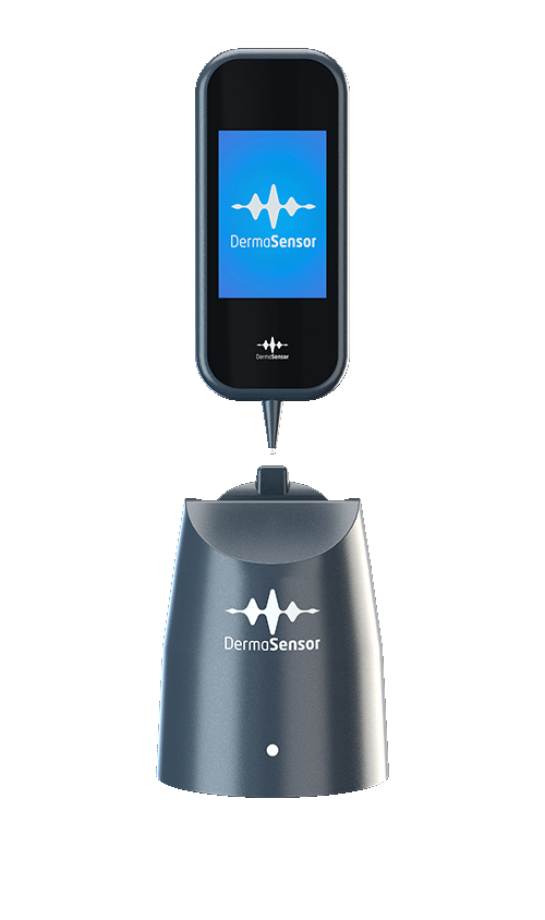
We now use DermaSensor — an FDA-cleared device that uses elastic scattering spectroscopy to analyze suspicious skin lesions in seconds. For patients aged 40 and above, it gives your provider an additional layer of clinical insight, helping determine whether a lesion warrants referral to a dermatologist.
DermaSensor is used alongside a full visual exam. It’s a tool that supports your provider’s clinical judgment, not a replacement for it.
After the five recordings, the device provides one of two results. Your provider will review this result alongside everything else they observed during your exam.

During your skin exam, any spot that looks suspicious is flagged for evaluation — not used as a general screening scan.

A small handheld tip is gently placed on the lesion, no needles or discomfort. Five readings take under a minute.

DermaSensor compares the lesion’s light-scattering properties to a database of malignant and benign lesion profiles.

The result guides your provider on whether to monitor the lesion or refer you to a dermatologist for further evaluation.
Early detection is key to treating various types of skin cancers before it has a chance to spread.
It’s especially important to get screened if you have a family history of skin cancer or other risk factors, such as fair skin, a lifestyle that exposes you to the sun often, a history of severe sunburn, or if you use tanning beds.
You should start getting regular or annual skin cancer screenings around age 20, although skin cancer can happen at any age.
The average age for a skin cancer diagnosis is around the age of 65, but melanoma in particular is one of the most common forms of cancer in younger people and should be taken seriously no matter what age you are.
Wearing sunscreen when outdoors is the best way to prevent skin cancer. Additionally, wear hats and long sleeves and limit your exposure when possible.
READY TO SCHEDULE YOUR NEXT APPOINTMENT?
NEW PATIENTS GET $100 OFF ANY COSMETIC TREATMENT
Take the first step towards your aesthetic goals with our complimentary 30-minute consultation. Simply fill out the form below and a patient care coordinator will be in touch shortly to help you schedule your consultation.
"*" indicates required fields
Take the first step towards your aesthetic goals with our complimentary 30-minute consultation. Simply fill out the form below and a patient care coordinator will be in touch shortly to help you schedule your consultation.
"*" indicates required fields
NEW PATIENTS GET $100 OFF ANY COSMETIC TREATMENT
Take the first step towards your aesthetic goals with our complimentary 30-minute consultation. Simply fill out the form below and a patient care coordinator will be in touch shortly to help you schedule your consultation.